Blood Vessels Labeled / Blood Vessel Wikipedia. The common cartoid artery extends from the brachiocephalic artery. Arteries, veins, and capillaries blood vessels flow blood throughout the body. Review the major systemic arteries of the body including those of the neck, arm, forearm, abdomen, pelvis, thigh, and leg in this interactive tutorial. Eventually, the smallest arteries, vessels called arterioles, further branch into tiny capillaries, where nutrients and wastes are exchanged, and then combine with other vessels that exit capillaries to form venules, small blood vessels that carry blood to a vein, a larger blood vessel that returns blood to the heart. The iliac, femoral, popliteal and tibial (calf) veins are the deep veins in the legs.
A primary purpose and significant role of the vasculature is its participation in oxygenating the body. Aside from capillaries, blood vessels are all made of three layers: There are five main types of blood vessels: The word vascular, meaning relating to the blood vessels, is derived from the latin vas, meaning vessel. Arteries, arterioles, capillaries, venules and veins.
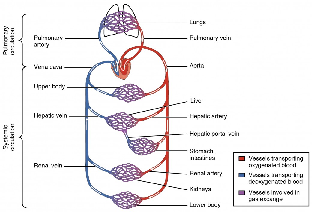
3 from Start studying blood vessels labeling. A blood vessel's main function is to transport blood around the body. The adventitia or outer layer which provides structural support and shape to the vessel The major veins in the A primary purpose and significant role of the vasculature is its participation in oxygenating the body. Anatomy of blood vessels review sheet 32 261 microscopic structure of the blood vessels 1. Veins (in blue) are the blood vessels that return blood to the heart. This occurs through two different circuits.
Start studying blood vessels labeling.
Free online quiz blood vessel labeling The function and structure of each segment of the peripheral vascular system vary depending on the organ it supplies. Best quiz blood vessel labeling; Its smooth surface decreases resistance to blood flow The iliac, femoral, popliteal and tibial (calf) veins are the deep veins in the legs. Bulky middle tunic contains smooth muscle and elastin 3. The word vascular, meaning relating to the blood vessels, is derived from the latin vas, meaning vessel. This article lists a series of labeled imaging anatomy cases by system and modality. Start studying blood vessels labeling. Review the major systemic arteries of the body including those of the neck, arm, forearm, abdomen, pelvis, thigh, and leg in this interactive tutorial. •formed where capillaries unite • extremely porous 1) venules: A capillary bed forms a maze of capillary vessels that lies between an arteriole and a venule. The superior vena cava is not labeled in figure 7.4.
To play this quiz, please finish editing it. When sphincter muscles are relaxed, the capillary bed is open, and blood flows through the capillaries. Blood vessels (labeled) coloring page. Very small branches that collect the blood from the various organs and parts are called venules, and they unite to form veins, which return the blood to the heart. Deep veins, located in the center of the leg near the leg bones, are enclosed by muscle.
Structure And Function Of Blood Vessels Anatomy And Physiology Ii from s3-us-west-2.amazonaws.com Blood vessel structure and function cardiovascular system: The microvasculature is composed of blood vessels that are smaller than 100 microns may only be seen through the microscope. The word vascular, meaning relating to the blood vessels, is derived from the latin vas, meaning vessel. Interactive physiology with quizzes cardiovascular system: The adventitia or outer layer which provides structural support and shape to the vessel Blood vessels are divided into two broad categories: Deoxygenated blood from the peripheral veins is transported back to the heart from capillaries, to venules, to veins, to the right side of the heart, and then. Arteries transport blood away from the heart.
The 4 valves are the aortic, pulmonary, mitral, and tricuspid valves.
The adventitia or outer layer which provides structural support and shape to the vessel There are five main types of blood vessels: The major veins in the Blood vessels (labeled) coloring page. Bonner walks through the dissection of a cat's blood vessels. Structure of blood vessel walls. The thick outermost layer of a vessel (tunica adventitia or tunica externa) is made of connective tissue. All blood vessels consist of a similar basic structure, which includes: This occurs through two different circuits. Blood is supplied to parts within the neck, head and brain through branches of the subclavian and common carotid arteries. Blood vessels consist of arteries, arterioles, capillaries, venules, and veins. Arteries (in red) are the blood vessels that deliver blood to the body. When sphincter muscles are relaxed, the capillary bed is open, and blood flows through the capillaries.
Learn vocabulary, terms, and more with flashcards, games, and other study tools. The iliac, femoral, popliteal and tibial (calf) veins are the deep veins in the legs. Related posts of the human blood vessels labeled digestive system worksheet answers. Aside from capillaries, blood vessels are all made of three layers: Arteries, arterioles, capillaries, venules and veins.
Blood Vessel Structure And Function Boundless Anatomy And Physiology from textimgs.s3.amazonaws.com Deep veins, located in the center of the leg near the leg bones, are enclosed by muscle. Veins return blood back toward the heart. This article lists a series of labeled imaging anatomy cases by system and modality. This occurs through two different circuits. The 4 valves are the aortic, pulmonary, mitral, and tricuspid valves. The microvasculature is composed of blood vessels that are smaller than 100 microns may only be seen through the microscope. The superior vena cava is not labeled in figure 7.4. Aside from capillaries, blood vessels are all made of three layers:
Veins return blood back toward the heart.
Inner layer is made of simple squamous. Blood vessels (labeled) coloring page. Figures 1 and 2 show the major arteries and veins of the body. Free online quiz blood vessel labeling Blood vessel labeling online quiz; Factors that affect blood pressure cardiovascular system: This occurs through two different circuits. The superior vena cava is not labeled in figure 7.4. The 4 valves are the aortic, pulmonary, mitral, and tricuspid valves. Arteries (in red) are the blood vessels that deliver blood to the body. Blood vessels are found throughout the body. Review the major systemic arteries of the body including those of the neck, arm, forearm, abdomen, pelvis, thigh, and leg in this interactive tutorial. The major veins in the